1999 | Dieffenbach-Medaille
Dieffenbach-Medaille Dr. med. J. C. Mustardé
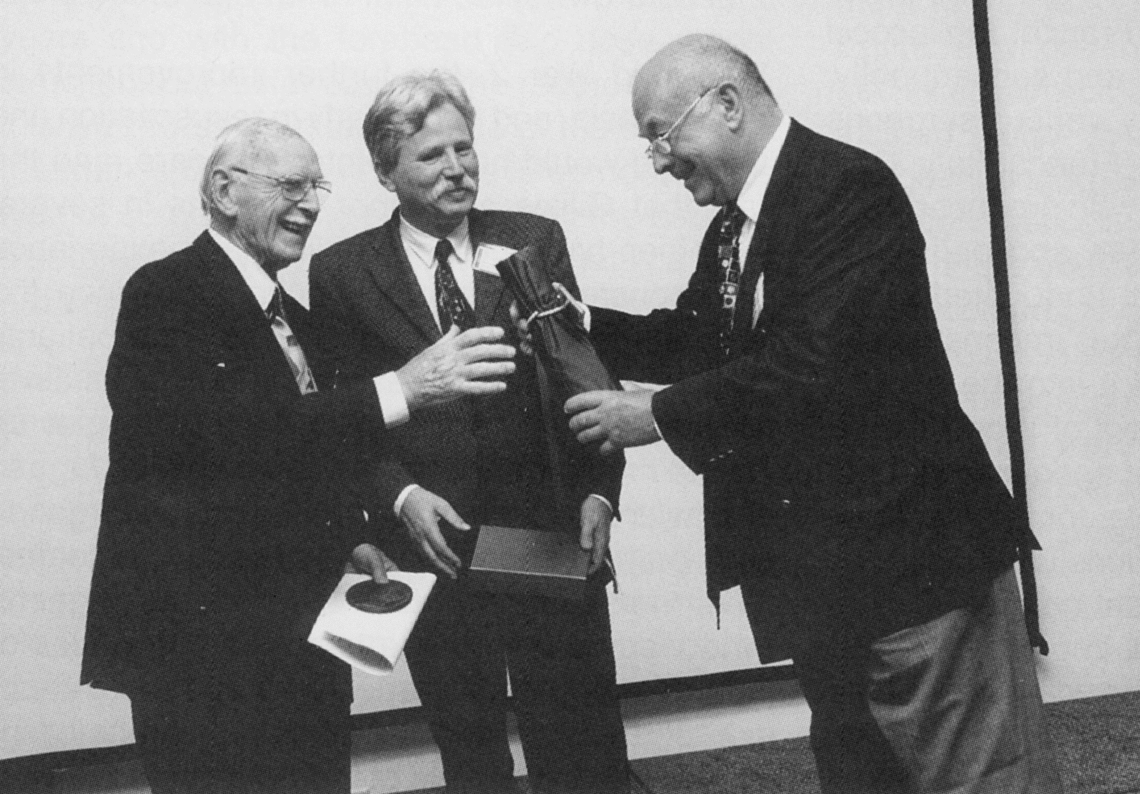
Die Dieffenbach-Medaille 1999 wurde Dr. med. J. C. Mustardé verliehen. Das Foto zeigt (von links): Dr. J. C. Mustardé, Prof. Dr. Michael Greulich und Prof. Dr. Neven Olivari.
„Dieffenbach-Vorlesung von Dr. J. C. Mustarde
Five years ago I had the honour of being invited to Berlin to take part in the commemoration of the 200th anniversary of the death of Dieffenbach, and it is therefore a special pleasure for me to have been honoured by the lnvitation of the German Plastic Surgery Association to come again to Germany to receive the Dieffenbach Award.
The name of Dieffenbach has been familiar to me for almost all of my fifty-eight years involvement with Plastic Surgery, and the Dieffenbach operation for advancing the cheek tissues to form the outer skin layer on which to reconstruct a lower eyelid, and which I illustrated in my Text book on Orbital Repair and Reconstructive Surgery, is the foundation of one of the basic techniques in use to this present time.
Dieffenbach was a surgeon with a very wide ranging experience, and his innovative writings on the whole field of Reconstructive Surgery were certainly an inspiration to others long after his death.
There were other writers on the subject, of course, even in the medieval times long before Dieffenbach’s time, Ambrose Pare in France, Tagliacocci in ltaly, with his nose repair and subsequently, particularly in the 19th century, various surgeons in different parts of Europe – France, ltaly, Germany, and in central Europe, who produced publications right through the 19th and early 20th Centuries. But the event that undoubtedly produced great innovations in this reconstructive type of surgery, and brought it into the public’s consciousness, was the advent of World War 1, when, with the introduction of ende-tracheal anaesthesia, surgeons were able, for the first time, to carry out facial reconstructions unhindered by a facial anaesthetic mask. I am no great historian, and I may be missing out important names, but for Britain and America the man who developed reconstructive surgery of the face more than any other, and published the first English text book shortly after the first world war about his experiences in this field was a New Zealander, .Sir Harold Gillies, working in Sidcup hospital in England.
His greatest contribution was the development of the infection-free method of transferring thick flaps of skin and underlying subcutaneous tissue from the abdomen to the face using a completely closed Tubed Pedicle Flap, brought up in stages – and which, by an incredible coincidence, was also claimed to have been developed during the same period, or slightly later, by a Russian surgeon named Filatov. (The flap was outlined and undermined, leaving attachments at either end, and was closed by sutures into a tube, the abdominal defect being closed direct. Two weeks later one end of the flap was divided and inserted on to a wrist – although in very big flaps the pedicle was only partly divided and complete division delayed for a further week. After two more weeks the remaining pedicle was divided and about half of the flap was inserted into the facial defect, followed after a further two weeks by the rest of the flap: 6-7 weeks in all.
By World War 2, the further improvements in anaesthesia, and particularly in resuscitation and what we would now call intensive care, and the fact that Gillies and other surgeons in several countries had been expanding their experience of reconstructive surgery techniques since the end of WW1, made it possible to keep patients with even the most grossly damaged faces alive, and specialist Maxillo-Facial units were set up by the medical services in the armies, as far as I am aware, of both sides, with general surgeons and oral surgeons working together in teams. With the passage of time the general surgeons who were part of these teams in the armies of both sides gradually advanced into reconstruction of areas other than the face; eventually involving the whole field of the body surface – and which later included the treatment of burns and their sequelae.
With the ending of the war this type of surgery developed very rapidly, with surgeons dedicated to work solely in this field, and this gave rise to the notion, certainly in the minds of the general public, that this type of surgery – which had been termed Plastic Surgery – was a completely modern concept. But nothing could be further from the truth …
When one thinks about it, invasive surgery within the body cavities – the abdomen, the thorax, and the skull (apart from trephining), has only been possible successfully in the last 130 years or so, and the only surgery which was possible before then, other than basic treatment of wounds, and possibly of some fractures, was on the outside of the body, involving skin and subcutaneous tissue – where people had at least a some knowledge of what they were looking at. lt is worth reminding ourselves that the first writings by Bilroth on abdominal surgery were published in 1881 – not quite 120 years ago.
The ability to carry out operations on the outside of the body, even including the use of some quite complex flaps, has existed for thousands of years, and we find records of surgical procedures on the surface of the body in ancient times, for example in Egypt almost three and a half thousand years ago, or in lndia some two thousand years ago with the forehead flap nose reconstruction. But as far back as the records go back this was surgery on the surface of the body, and the truth is that it is only the development of this type of surgery on the outer parts of the body as a surgical specialty in its own right that is a modern concept.
Dieffenbach may not have been the first to try and systematise the principles of reconstructive surgery of his day, but his contribution, with many procedures not recorded before, certainly highlighted and developed this type of surgery to a stage considerably above what had been available until then, and he must without doubt be regarded as one of the founders of Plastic Surgery, at least as we knew it in the earlier half of this century.
There is no doubt, however, that, as in every branch of surgery, the discoveries and innova tions which have taken place in reconstructive plastic surgery in the latter part of this century have brought forward the rate of development of what can now be achieved to such an extent that those who were not born till after the middle of the century could not be blamed for thinking that Plastic Surgery only began in the lifetime perhaps of their fathers.
When we consider some of the advances which have taken place in the last few decades alone, in particular the advent of micro vascular techniques, and the new approaches which are now available for the non-invasive investigative and operative techniques of almost any part of the body, the 3D scanning of the inside the skull and the host of modern day aids which are available to today’s plastic surgeon, it may be of interest to the younger generation of surgeons to glimpse a little of the State of the Art in Plastic Surgery in the far off days when people like myself began working in this field.
My own involvement in reconstructive surgery began in WW2, when I had enlisted in the Medical Corps of the British army in 1940. In that very cold winter – with the genuine intention of increasing the very restricted rationed diet of my meng – I illegally shot a few deer in the Highlands of Scotland, and I was posted as a punishment to the war zone in the Western Desert in Egypt. – where they probably expected me to get killed … Actually I didn’t get killed. – and instead found myself working as assistant to a famous London Surgeon Henry Stallard, who, although basically an ophthalmic surgeon, had published a book before the war about repairing and reconstructing damaged eyelids, and other orbital injuries. All the orbital injuries, not only of our own troops, but also German and ltalian Prisoners of War, were sent direct to Stallard’s wards in Cairo. We operated almost every day, and this work of reconstruction absolutely fascinated me.
As a small boy I had always liked repairing things and inventing things, and this type of surgery, restoring and building up eyelids and orbital stuctures, made me realise that that was exactly what I wanted to do in life.
The main problem with reconstructive work in those days was the constant threat of infection, particularly in wounds where the surrounding tissue was badly traumatised, and in wounds like that maggots, the larvae of the big blue flies, which were often beginning to feed on the necrotic tissue in the infected wounds were regarded very favourably – an attitude which I note is today being rediscovered!
There were no antibiotics and in the battle against infection reliance was mainly placed on using disinfectant solutions, particularly chlorinated Eusol. Just before the war, however, following on the discovery by the German bacteriologist Gerhard Domagk, working with Bayer, that the coloured dye Prontosil Red, could kill bacteria without damaging the tissues, the active principle Sulphanilamide, was isolated as the first of the Sulpha bacteriocidal drugs – mostly used topically as a powder.
They were moderately successful, but extremely expensive, and in hot places like Egypt they were less successful; and more importantly, were no help at all in desert areas against the deeply spreading skin ulceration caused by diptheria organisms.
At about the same time, the accidental discovery by a fellow Scot, Alexander Fleming, of the incredible bacterial inhibiting action of Penicillin, the first antibiotic, had taken place, and its potential was only now being explored experimentally. By 1941 it had become available in small quantities to hospitals and was hailed as a miracle drug. Unfortunately supplies were extremely limited and by 1942, when I was working in the Lybian port of Tobruk, only small quantities were available to the army for life and death situations, and not for surface surgery.
(On this matter of wound infection, it is interesting that when, in June 1942, Tobruk was surrounded by Rommel – and eventually captured the water supply got blown up by some English idiot, acting prematurely on the order to destroy everything that the enemy could use if the town was captured. And in the hospital, where we were operating literally night and day to deal with the increasing numbers of wounded, we had to go on operating using the same water for 48 hours, and it became so filthy that we had to filter it through a wet towel, but the infection rate did not alter as far as we could determine.)
Although the tubed pedicle flap was available for transfer of thick, bulky flaps, and had greatly reduced the amount of infection in reconstructive work where direct open transfer flaps such as the cross leg and other open flaps, invariably became infected at the base, they were too slow for use in a war situation where beds were in very short supply, taking anything up to 7 weeks to complete the transfer, and many efforts were made to find a way to shorten the rate of flap transfer and yet avoid the risk of infection.
I had myself developed a technique to cut down the total transfer time to 21 days, and subsequently published a paper on The Rapid Flap Technique in the American Plastic and Reconstructive Surgery Journal. The whole flap was undermined at the first stage, with lateral pedicles being retained, and the under surface of the flap, as well as the raw surface of the donor site was temporarily covered with split skin grafts. One end of the flap was divided and inserted on the wrist at the first operation, and the lateral. The lateral pedicles were divided during the first week so that the flap was ready to have its remaining base divided at the end of the first week and the terminal half inserted in the defect. At the end of the second week the wrist attachment was freed and the whole flap was then put in place.
lt had a measure of success, but more or less involved tying the patient to the bed in case he pulled the flap off, but the ending of the war mean there was not the same problem about time, and also, working as I was in Sir Harold Gillies Unit it seemed wiser for me to use his tubed pedicle flap if I wanted to keep my job!
Using comparatively long flaps with a ratio of 3, or sometimes 4, to 1, with pedicles which we hoped contained enough arteries and veins to keep the blood circulation functioning during the transfer, we were very much concerned with resuscitation of flaps which were not doing well. We appreciated that a blue flap was a congested flap with poor venous drainage, while a white flap was an ischaemic flap with poor arterial supply, and a great deal of argument went on as to the best way to manage the problem of a potentially dying flap.
With blue flaps, some advocated intermittent massage of the flap to physically empty the veins of blood, a performance that was carried out for
24 hours or more by the junior assistants. Other believed that application of moderate warmth would get the blood flowing more freely. Leeches were advocated to drain off the stagnant blood (and it is interesting to learn of their use being brought back again as if it was a new discovery). White flaps, also produced vehement arguments, this time between those who considered the correct procedure was to apply ice cold compresses to slow down metabolism because of lowered available oxygen, while the opposition advocated applying warmth to encourage the vessels to dilate.
I am not sure that what we did really made much difference as many of these doubtful flaps died whatever was done to try and save them. The real problem, of course, was that we did not have an adequate knowledge of the peripheral blood circulation which would have ensured that the flaps were always designed in a manner which made sure the skin circulation had adequate vascular vessels incorporated in it.
Microvascular surgery had not even been thought of at that time, but we sometimes discussed the possibility of dissecting out a section of a small vein from the dorsum of the foot and using it to connect up the congested veins in the flap to a vein in the recipient site. But I never heard of anyone actually doing this, and we were always concerned that using small veins as free grafts like this would result in their thrombosing, with the possibility of emboli. The smallness of the arteries, even if we had had a microscope, would have been impossible to link up securely with the needles and materials then at our disposal.
With regard to split skin grafts, which at first we learned to cut very expertly free hand with an open razor, and later with a roler-guarded Blair Type knife, not very much was known in the early days about homograft skin graft incompatability. Professor Medawar and our own Tom Gibson in Scotland had just begun their historic research into skin compatability and the phenomenon of rejection, and the possibility of finding individuals, other than identical twins, whose skin might not be rejected was often disussed.
By this time I had returned to UK as a repatriated Prisoner of War, and was temporarily posted to an army camp. The question of whether blood groups might be a factor in determining if one person’s skin could survive on another was much in discussion, and I got permission to ask for volunteers (with the promise of a free beer) to Iet me try out an experiment using 50 individuals, each giving a small Thiersch graft type of skin so that we could try out various combinations of skin transfers according to blood groups.
Two of the soldiers queing to have qa graft resected fainted and fell on the floor when the local anaesthetic was injected, and one of them split his forehead skin and had to have sutures inserted – which called for an official Army enquiry Iasting four hours! And of course, none of the homografts survived permanently, although we noticed that some of the grafts survived Ionger than others, but we deuced that rejection did not seem to have any relation to the blood groups.
Although we used stainles steelwire extensively at jaw fracture sites (mini-plates had not then been invented), it was always regarded as taking a risk to bury foreign material, metal, Bakelite – a proteinbased substance similar to modern plastics (which did not exist in the early days), and other inert materials beneath the skin because of what was regarded as a considerable risk that the foreign body would become the focus for an abcess, or would even be extruded. And one of the absolutely forbidden procedures was to leave a foreign substance protruding through the skin or mucosal covering layer, whether in the eye socket, or the mouth, or on any other part of the body. Nobody did this deliberately – although from time to time there were cases reported where a glass sphere buried in an eyesocket, or some other foreign substance in some other part of the body, had become exposed without being rejected. But on principle it was regarded as unsurgical to do this deliberately, and we went to great lengths to avoid the possibility.
I still don’t know how intra osseous dental implants do not give rise to osteo myelitis of the underlying bone, or how the semi-buried orbital implants into which the posterior projection of an artificial eye can be inserted to give mobility to the overlying prothesis, succeed in most instances. Perhaps it was simply that we just did not have enough courage.
Looking back as I have briefly done just now, of all today’s advances that I would most have wanted to have I would regard micro-vascular surgery, with the development of the free flap, and particularly the composite free flap including muscle and even bone, as one of the greatest advances to have been made in Plastic Surgery since the fact that a small pinch of skin could be snipped off and could survive and grow on a raw surface of the same individual was discovered by the Reverdin over a century ago.
Had advances of that nature been available to him, I wonder what Dieffenbach himself would have been able to do in his quest to help mankind. And with this in mind I salute him as one of the truly great figures in the history of the development of today’s Reconstructive Plastic Surgery.“
Quelle: Mitteilungen VDPC, Nummer 11, 6. Jahrgang Dezember 1999